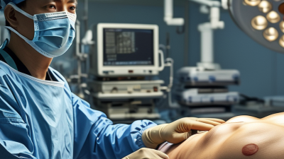
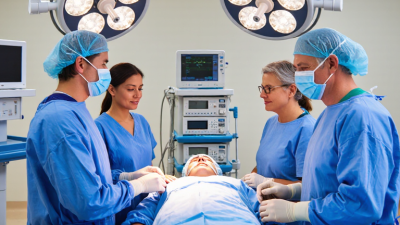
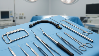
Bone Reconstruction describes the surgical restoration of damaged, missing, or poorly formed bone. It may follow severe injury, infection, tumor removal, congenital conditions, or failed healing. Surgeons assess the defect’s size, location, blood supply, and the patient’s overall health before choosing a reconstruction plan. The aim is not simply to fill a gap. The new structure must also support healing and, where needed, restore function.
The scale of bone grafting is significant. In a 2005 review in Injury, orthopedic trauma specialist Panayiotis V. Giannoudis and colleagues wrote that bone grafting was “the second most common tissue transplantation worldwide,” estimating more than two million procedures annually. That figure is historical, not a current annual count, but it shows the treatment’s established role. Reconstruction may use a patient’s own bone, donor bone, or engineered materials. Each option has trade-offs. No single method fits every defect.
In practical terms, reconstruction can involve shaping a graft, securing it with plates or screws, and monitoring healing through follow-up imaging. The body must connect the graft with living bone. That takes time. Outcomes depend on factors such as blood flow, infection control, smoking, and the defect’s complexity. Even careful surgery cannot guarantee identical healing for every patient. This guide explains how Bone Reconstruction works, what materials and techniques may be considered, and which questions patients can discuss with an orthopedic specialist.
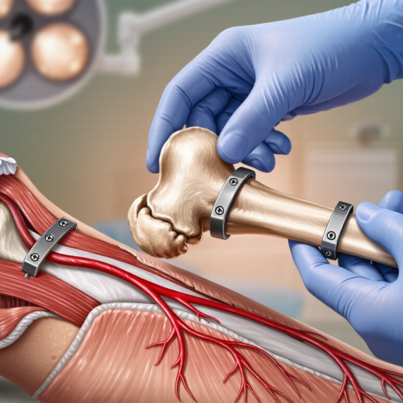
Understanding Bone Reconstruction Bone reconstruction means rebuilding or reshaping bone when its structure has been lost, weakened, or formed differently. It may follow a severe fracture, removal of diseased tissue, a birth difference, or bone loss around the jaw. The aim is practical: restore support, alignment, or function, not simply make an image look normal. That distinction matters. Reconstructed bone may not feel or perform exactly like the original.
Treatment depends on the bone’s location, the size of the gap, blood supply, and overall health. A surgeon may stabilize existing bone with plates or screws, or fill a defect with a graft. Grafts can come from the patient, a donor, or synthetic material. Over time, living bone may grow into or around the graft. Healing can take months, and results are not always predictable. Even “reconstruction” can describe different procedures, so ask what it means in a specific case.
Tips: Ask which bone is affected, what material may be used, and how healing will be checked. Request a plain-language explanation of risks and alternatives. Bring a list of medicines and previous operations to appointments. Small details help.
Bone reconstruction restores or reshapes bone that is too thin, weak, or damaged to support normal function. It may be considered after an injury, infection, surgery, or gradual bone loss. The reasons vary. In the jaw, for example, bone can shrink after a tooth is lost, leaving too little support for some dental treatments. A person may also need reconstruction after removal of a growth or treatment that affected nearby bone.
The right approach depends on the cause, location, and amount of bone loss. A clinician may review medical history and use X-rays or other imaging to assess the area. Treatment can involve moving existing bone or using a graft, but reconstruction is not always necessary. Healing may take time, and results differ between people. Grafts can fail to integrate, and infection or delayed healing can occur. That deserves an honest discussion before treatment.
Tips: Ask what problem reconstruction is meant to solve, what alternatives exist, and how healing will be monitored. Share medications, health conditions, and smoking history with your care team. Bring questions to the appointment; it is easy to forget them.
Choosing a bone reconstruction method depends on the missing bone’s size, location, and cause. A small gap in a stable area may need a different approach from a large defect near a joint. Surgeons also assess blood supply, surrounding muscle and skin, infection risk, and the patient’s overall health. X-rays or CT scans show the shape of the defect; examination helps assess pain and function. No scan tells the whole story.
Options may include grafting, bone transport, or a graft with its own blood supply. A graft can come from the patient, a donor, or a synthetic material. Bone transport gradually moves healthy bone across a gap, but treatment can take months and requires close follow-up. The best fit depends on healing potential, previous operations, and the function the patient hopes to regain. Plans may change if the tissues heal differently than expected. That uncertainty deserves an honest discussion.
Tips: Ask why the proposed method suits your specific defect, how long healing may take, and what follow-up involves. Bring prior imaging and a list of medicines. Ask about likely limitations, too. Those details can be easy to overlook.
The Main Steps of a Bone Reconstruction Procedure
Bone reconstruction repairs or rebuilds an area of bone that is missing, damaged, or too thin for its intended function. The exact procedure depends on the injury, the person’s health, and the location of the bone. Planning begins with a medical history, a physical examination, and imaging such as X-rays or a CT scan. These images help the surgical team assess the bone’s shape and choose an approach. Still, scans cannot answer everything.
Before surgery, the clinician explains anesthesia, likely risks, and expected recovery. During the procedure, the surgeon reaches the affected area through an incision and prepares the bone. A graft may come from the patient, a donor, or a processed material. The surgeon positions it where new bone growth is needed, then may secure it with plates, screws, or other supports. Small details matter. A graft that shifts can heal poorly, so stability is checked before the incision is closed.
Afterward, the care team monitors pain, swelling, wound healing, and signs of infection. Follow-up visits and imaging help show whether the reconstruction is integrating. Healing often takes months, not days, and activity may need to be limited while the bone strengthens. Recovery varies; progress can be uneven. Even a well-planned procedure does not guarantee a particular result, so patients should ask their clinician which warning signs need prompt attention.
| Step | What Happens | Purpose | Typical Considerations |
|---|---|---|---|
| 1. Assessment and diagnosis | The care team reviews medical history, examines the affected area, and may use X-rays, CT scans, or other imaging. | To define the size and location of the bone defect and identify factors that could affect healing. | The plan depends on the bone involved, the cause of bone loss, overall health, and whether infection or soft-tissue damage is present. |
| 2. Treatment planning | The surgeon selects a reconstruction approach, such as a bone graft, a bone substitute, bone transport, or a combination of methods. | To choose an approach suited to the defect and the patient’s treatment goals. | Graft material may come from the patient, a donor, or a manufactured substitute. Not every method is appropriate for every defect. |
| 3. Anesthesia and preparation | Anesthesia is provided, and the surgical area is cleaned and prepared. The type of anesthesia depends on the procedure and patient. | To support patient comfort and reduce the risk of contamination during surgery. | Preoperative instructions may include medication guidance, fasting, and arrangements for recovery after the procedure. |
| 4. Access and site preparation | The surgeon reaches the affected bone and prepares the area. Damaged tissue may be removed, and the defect may be shaped for reconstruction. | To create a suitable site for the graft or other reconstruction method. | If infection is present, it may need to be controlled before or as part of reconstruction. |
| 5. Bone reconstruction | Graft material or another reconstruction method is placed at the defect. In some cases, bone is gradually lengthened or transported over time. | To restore bone continuity, support, shape, or length and encourage new bone formation. | The technique varies. Some procedures use a graft alone; others use a device or fixation system to support the reconstruction. |
| 6. Stabilization and closure | The reconstructed area may be secured with plates, screws, rods, an external frame, or other fixation. The incision is then closed and dressed. | To keep the bone aligned and stable while healing begins. | Weight-bearing or movement restrictions may be prescribed, depending on the bone and the fixation used. |
| 7. Recovery and follow-up | The care team monitors the wound, symptoms, and bone healing. Follow-up visits and imaging may be used to assess progress. | To identify complications and check whether the reconstruction is healing as planned. | Healing commonly takes weeks to months and varies by procedure, bone, health, and other factors. Rehabilitation may be recommended. |
Recovery after bone reconstruction is gradual, and the timeline depends on the bone, graft type, and overall health. Expect swelling, bruising, and soreness near the surgical site during early healing. Your care team may recommend limited weight-bearing, wound checks, and follow-up imaging. AAOS guidance notes that many fractures take about 6–8 weeks to heal, but reconstructed bone can require longer. That figure is only a reference, not a personal deadline. Healing is rarely a straight line; a quiet week can still be followed by discomfort.
A 2016 BMJ Open systematic review examined 40 studies involving 7,516 procedures. It found smokers had 2.2 times the risk of delayed or failed bone healing, with healing taking an average of 27.7 days longer. This does not predict an individual outcome, but it shows why smoking cessation matters. Follow your surgeon’s instructions on activity, medication, and nutrition. Contact the care team about worsening pain, fever, increasing redness, or drainage.
Tips: Keep appointments, use mobility aids as directed, and record changes in pain or swelling. Don’t rush activity because you feel better; ask before increasing weight or exercise. Recovery can feel slow. That deserves honest discussion with your clinician.