Spinal Implants are becoming central to modern spine reconstruction, deformity correction, and motion preservation. The market is expanding, but growth alone does not prove clinical value. Fortune Business Insights estimated the global spinal implants market at approximately USD 10 billion in 2023, with continued growth expected through 2030. Grand View Research reports similar momentum, driven by aging populations, degenerative disc disease, and improved surgical technology.
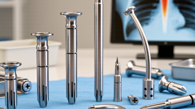
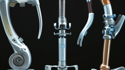
This article examines ten major types, including interbody cages, pedicle screws, spinal rods, anterior plates, artificial discs, expandable cages, vertebral body replacements, spinal hooks, sacroiliac fusion implants, and bone-growth technologies. Each device serves a different mechanical purpose. Some restore height. Others stabilize unstable segments. A few aim to preserve movement. The operating room makes these differences tangible: titanium screws anchor into vertebrae, while cages support a compressed disc space.
Clinical judgment remains more important than product novelty. Spine surgeon Dr. Alexander R. Vaccaro has emphasized, “Patient selection is the key to success.” That principle deserves attention. The most advanced implant may still be unsuitable for a specific patient. The FDA also stresses that spinal devices require careful consideration of benefits, risks, indications, and long-term follow-up.
This overview uses clinical literature, regulatory guidance, and industry reports to explain how these implants differ. However, market statistics can hide uneven outcomes. Surgical success depends on anatomy, diagnosis, technique, bone quality, and rehabilitation. Readers should treat this guide as an informed starting point, not a substitute for advice from a qualified spine specialist.
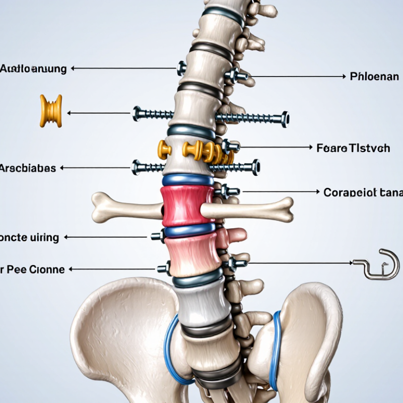
Spinal implants are designed to restore stability, alignment, or movement after injury, degeneration, or deformity. The ten common categories include interbody cages, pedicle screws, rods, spinal plates, hooks, artificial discs, vertebral body replacements, kyphoplasty systems, spinous-process devices, and sacroiliac fusion implants. Each serves a different mechanical purpose. A cage supports the disc space and encourages bone growth. Screws and rods create a controlled frame. An artificial disc aims to preserve motion instead of creating fusion.
Design decisions depend on anatomy, bone quality, surgical access, and the patient’s daily demands. Materials may include titanium alloys, cobalt-chromium, or radiolucent polymers. Surface texture also matters because it can support bone attachment. A 2024 report from Grand View Research valued the global spinal implants market at more than 10 billion U.S. dollars, reflecting substantial clinical use and continuing design development. Market size is not proof that one implant works best.
Clinical evidence requires restraint. The AHRQ Healthcare Cost and Utilization Project recorded about 465,000 spinal fusion hospital stays in the United States during 2014. That figure shows demand, but not success in every patient. A device can look precise on imaging yet fail to relieve pain. Surgeons therefore assess neurological findings, imaging, bone density, and long-term risks before choosing an implant. No design fits every spine. Patient expectations can also be imperfect. Clear follow-up remains essential, especially when fusion, loosening, or adjacent-segment stress is possible.
What Are the Top 10 Types of Spinal Implants?
The ten common types include interbody cages, artificial discs, pedicle screws, rods, plates, hooks, wires, spacers, vertebral body replacements, and sacroiliac joint implants. Interbody cages restore disc height and support fusion between vertebrae. They may use titanium, polyether ether ketone, or porous materials. Their design affects bone growth, positioning, and imaging quality.
Artificial disc replacement implants aim to preserve motion in selected patients with painful disc degeneration. They replace a damaged disc with endplates and a mobile core. Clinical selection remains critical. Patients usually need controlled symptoms, suitable anatomy, and limited facet-joint disease. A 2024 Grand View Research market assessment projected approximately 5% annual growth for the spinal implants market through 2030. This reflects expanding surgical demand, not proof that every implant improves outcomes. The North American Spine Society emphasizes imaging, neurological findings, and conservative treatment history before surgery. Outcomes vary.
Tips: Ask how the implant supports your specific anatomy. Request evidence from long-term studies. Confirm whether bone quality, alignment, and facet health affect eligibility. Do not assume newer means better. That assumption deserves scrutiny.
| No. | Spinal Implant Type | Common Materials | Primary Purpose | Typical Spinal Region or Approach | Key Design Features | Limitations and Considerations |
|---|---|---|---|---|---|---|
| 1 | Interbody Fusion Cage | PEEK, porous titanium, titanium alloy, or structural allograft | Restores disc-space height, supports vertebral endplates, and promotes fusion between adjacent vertebrae. | Cervical, thoracic, or lumbar spine; used through anterior, lateral, oblique, or posterior approaches. | Hollow or fenestrated body for bone graft placement; teeth, ridges, or textured surfaces may improve initial stability; some designs provide lordotic or angled correction. | Requires successful bone fusion; subsidence, migration, nonunion, and adjacent-segment degeneration may occur. Proper sizing and endplate preparation are important. |
| 2 | Cervical Interbody Device | PEEK, titanium alloy, porous titanium, or allograft bone | Replaces a diseased cervical disc after discectomy and helps restore cervical disc height and alignment. | Anterior cervical spine, most commonly between the vertebral bodies. | Compact footprint, graft chamber, endplate-contact surfaces, and optional integrated fixation features such as screws or teeth. | May be associated with subsidence, nonunion, dysphagia related to the surgical approach, or implant migration. It eliminates motion at the treated level when used for fusion. |
| 3 | Lumbar Posterior Interbody Cage | PEEK, titanium alloy, porous titanium, or carbon-fiber-reinforced polymer | Provides anterior column support and a fusion surface while allowing decompression of lumbar nerve roots. | Posterior lumbar spine; commonly used in PLIF or TLIF procedures. | Expandable or fixed-height options; angled profiles; graft windows; and surfaces designed to resist migration. | Posterior approaches may involve nerve-root manipulation and disruption of posterior spinal structures. Risks include subsidence, cage migration, dural injury, and pseudarthrosis. |
| 4 | Lateral or Oblique Interbody Cage | PEEK, titanium alloy, or porous titanium | Restores disc height, improves foraminal dimensions, and supports indirect decompression and spinal alignment correction. | Lumbar spine through lateral, extreme-lateral, or oblique anterior-to-psoas approaches. | Wide footprint spanning stronger regions of the vertebral endplates; often available in multiple heights, widths, and lordotic angles. | Approach-specific risks may include vascular injury, psoas or lumbar plexus irritation, thigh symptoms, subsidence, and difficulty treating certain anatomy. |
| 5 | Artificial Cervical Disc Replacement Implant | Cobalt-chromium, titanium alloy, stainless steel, ceramic, polyethylene, or combinations of these materials | Replaces a diseased cervical disc while preserving motion at the treated level. | Anterior cervical spine, positioned between adjacent vertebral bodies. | Mobile core or articulating surfaces; endplate fixation features; radiopaque markers; and designs intended to preserve segmental movement. | Not appropriate for every patient, particularly those with severe facet disease, instability, osteoporosis, infection, or significant deformity. Heterotopic ossification, wear, migration, and revision surgery are possible. |
| 6 | Artificial Lumbar Disc Replacement Implant | Metal alloys, polyethylene, ceramic, or combinations of metal and polymer components | Replaces a symptomatic lumbar disc and aims to maintain motion while reducing discogenic pain in carefully selected patients. | Anterior lumbar spine, usually through an anterior retroperitoneal approach. | Mobile bearing or articulating core, vertebral endplate components, fixation teeth or keels, and radiographic markers for positioning. | Requires suitable facet joints, bone quality, alignment, and vascular anatomy. Potential concerns include implant wear, migration, subsidence, heterotopic ossification, and vascular or approach-related complications. |
| 7 | Pedicle Screw and Rod System | Titanium alloy, cobalt-chromium alloy, or stainless steel | Provides posterior fixation, controls spinal motion, supports correction, and stabilizes the spine during fusion. | Most commonly used in the thoracic and lumbar spine; selected cervical applications are also possible. | Screws anchor into vertebral pedicles and connect to longitudinal rods using fixed or polyaxial screw heads; cross-links and reduction features may be included. | Malposition can affect neural or vascular structures. Other risks include screw loosening, rod fracture, adjacent-level stress, infection, and nonunion. |
| 8 | Anterior Cervical Plate and Screw System | Titanium alloy, stainless steel, or radiolucent polymer components with metallic screws | Adds anterior stability after cervical discectomy or corpectomy and helps maintain alignment during fusion. | Anterior cervical spine, placed over the front of the vertebral bodies and interbody graft or cage. | Low-profile plate, locking or constrained screws, variable-angle fixation, and radiographic markers for implant assessment. | Potential complications include dysphagia, screw loosening or back-out, plate prominence, adjacent-level ossification, and difficulty with revision exposure. |
| 9 | Spinal Hooks and Sublaminar Fixation System | Titanium alloy, cobalt-chromium alloy, or stainless steel | Provides posterior fixation when pedicle screw placement is unsuitable or when additional segmental control is needed. | Thoracic and lumbar spine; may be used in deformity correction and selected revision procedures. | Hooks engage the lamina, transverse process, or other posterior elements and connect to rods or bands for construct stability. | Fixation strength depends on bone quality and posterior-element anatomy. Placement may risk dural or neural structures, and hooks may provide less three-column fixation than appropriately placed pedicle screws. |
| 10 | Vertebral Body Replacement Cage | Titanium alloy, porous titanium, or expandable metal implants | Reconstructs the anterior spinal column after corpectomy caused by tumor, trauma, infection, or severe vertebral-body collapse. | Cervical, thoracic, or lumbar spine; used through anterior, lateral, or posterior-assisted approaches depending on the case. | Fixed or expandable height, endplate-contact surfaces, central graft chamber, and the ability to restore vertebral-body height and alignment. | Often requires supplemental fixation. Risks include migration, subsidence, hardware failure, nonunion, and complications related to the underlying disease or surgical approach. |
Spinal implants support stability when bones, discs, or ligaments cannot provide enough support. The ten common types include:
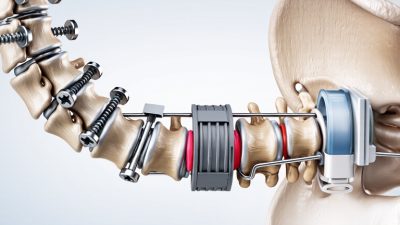
Selection depends on spinal level, bone quality, curvature, nerve pressure, and the planned surgical approach. A surgeon may combine screws and rods for lumbar stabilization. Cervical procedures may use smaller plates, screws, or specialized hooks. Cross-links can improve rotational control, but they are not required in every construct. No implant is universally best. That point is easy to overlook. Imaging, physical examination, and the patient’s healing capacity should guide decisions. Even experienced teams may revise the plan during surgery when bone is softer than expected.
Tips:
Ask which implant supports the main surgical goal. Confirm whether fusion is planned and where it will occur. Discuss possible movement limits after surgery. Ask how implant position will be checked using imaging. Bring questions about bone health, smoking, medications, and previous operations. Clear answers matter. They often reveal whether the proposed fixation system fits your anatomy and recovery needs.
What Are the Top 10 Types of Spinal Implants?
Vertebral body replacement implants are designed for severe spinal damage caused by tumors, fractures, or infection. They remove the unstable vertebral body and restore structural support. Expandable cage implants can then open gradually inside the spine. This helps surgeons adjust height during reconstruction. The goal is practical: restore alignment, protect neural structures, and create a stable platform for fusion.
The market is expanding. Grand View Research estimated the global spinal implants market at about USD 11.8 billion in 2023, with continued growth projected through 2030. Clinical evidence is more cautious. A 2023 systematic review in Global Spine Journal reported improvements in pain and vertebral height after expandable cage reconstruction, but study quality varied. That matters. A larger implant is not automatically a better implant. Patient bone quality, tumor location, spinal level, and remaining stability must guide selection. Surgeons also assess endplate damage and the risk of cage subsidence.
Tips: Ask whether a fixed or expandable cage fits the defect. Request details about load sharing, fixation, and fusion plans. Imaging should confirm alignment after surgery. Patients with weak bone may need additional support. I would not treat market growth as proof of clinical superiority. Long-term comparative evidence still needs strengthening.
Spinal implants are selected according to the affected spinal region, the type of instability or deformity, and the surgical objective. The chart shows the typical number of vertebral levels involved in common implant applications; actual constructs vary by patient and procedure.
Vertebral body replacement and expandable cage implants are primarily used to reconstruct a vertebral column defect after tumor removal, trauma, infection, or severe collapse. Other implant categories support fusion, stabilization, motion preservation, or deformity correction.
Kyphoplasty and vertebroplasty address different problems from deformity-correction implants. Vertebroplasty injects bone cement into a fractured vertebral body. Kyphoplasty first creates a cavity with a balloon. Cement then fills the space. The balloon may restore some vertebral height, but results vary.
Doctors review pain patterns, imaging, bone quality, and neurological findings before recommending either procedure. Important risks include cement leakage, infection, nerve irritation, and adjacent-level fractures. Careful needle placement and real-time imaging reduce, but never remove, these risks.
Deformity surgery uses a broader implant system. Common categories include pedicle screws, spinal rods, hooks, interbody cages, expandable vertebral body implants, osteotomy fixation devices, and sacropelvic screws. These components help correct scoliosis, kyphosis, instability, or vertebral collapse. Interbody cages can restore disc height and support bone fusion. Rods and screws maintain alignment while healing occurs.
A rigid construct may improve correction but can increase stress at nearby levels. An expandable implant may support a damaged vertebra, yet it requires precise sizing and placement. Bone density, spinal flexibility, age, and previous surgery also influence implant selection. Surgical planning often combines standing X-rays, computed tomography, and magnetic resonance imaging. Even experienced teams sometimes revise the plan during surgery when bone quality or anatomy differs from scans. This uncertainty deserves honest discussion.